UCSF Thoracic Imaging 2023
$35,00
Samples for Courses Can be found here : Free Samples Here!
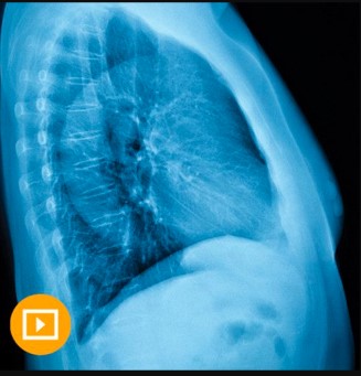
UCSF Thoracic Imaging 2023 This online CME course offers a practical approach to imaging of the pulmonary, vascular, and cardiac systems. It’s designed as a comprehensive review for radiologists who interpret thoracic imaging studies, focusing on the diverse use of CT, including PET, MRI, and radiography correlations.
UCSF Thoracic Imaging 2023
Radiology CME: The Latest in Thoracic Imaging
This online CME course offers a practical approach to imaging of the pulmonary, vascular, and cardiac systems. It’s designed as a comprehensive review for radiologists who interpret thoracic imaging studies, focusing on the diverse use of CT, including PET, MRI, and radiography correlations.
Concise continuing medical education lectures span a broad cross-section of thoracic topics and emphasize recent updates of established clinical guidelines, including lung cancer screening, management of pulmonary nodules, and a review of high-resolution CT of the lung in the context of updated diagnostic criteria for interstitial pneumonias.
UCSF Thoracic Imaging speakers also discuss the synthesis of cardiac and pulmonary findings in patients presenting with acute symptoms (i.e. pulmonary embolism, acute aortic syndromes, cardiac disease), and on post-operative patients.
Accreditation
The University of California, San Francisco School of Medicine (UCSF) is accredited by the Accreditation Council for Continuing Medical Education (ACCME) to provide continuing medical education for physicians.
Designation
UCSF designates this educational activity for a maximum of 12.25 AMA PRA Category 1 Credits™. Physicians should claim only the credit commensurate with the extent of their participation in the activity.
The total credits are inclusive of 13.00 in CT and 2.00 in Plain Film.
Date of Original Release: May 8, 2023
Series Expiration Date: May 7, 2026 (deadline to register for credit)
Estimated Time to Complete Activity: 12.25 hours
CME credit is obtained upon successful completion of an activity post-test and evaluation. CME Credit registration forms must be submitted prior to series expiration date. Certificates will be dated upon receipt and cannot be dated retroactively.
Learning Objectives
At the conclusion of this activity, the participant will be able to:
- Interpret high-resolution CT scans of the lungs and provide focused differential diagnoses
- Apply a practical approach to the imaging of lung infections
- Identify typical manifestations of acute and chronic aortic diseases
- Utilize cross-sectional imaging in the evaluation of pulmonary embolism
- Differentiate benign vs. malignant lung nodules/masses on CT and PET
- Implement the use of CT and PET in the evaluation and staging of lung cancer
Intended Audience
Radiologists, physician assistants, and nurses.
Topics:
Diffuse Lung Disease and Chest Radiographs
- Multi-disciplinary Approach to Diffuse Lung Disease – Brett M. Elicker, MD
- An Approach to Distinguishing Fibrotic Lung Diseases – Maya Vella, MD
- Cystic Lung Disease – Kimberly G. Kallianos, MD
- Mosaic Attenuation-Perfusion – Brett M. Elicker, MD
- Airways Diseases – Maya Vella, MD
- Chronic Consolidation – Brian M. Haas, MD
- Challenging Chest Radiographs – Maya Vella, MD
- Iatrogenesis Imperfecta – Post‐Op Findings in the Chest – Kimberly G. Kallianos, MD
- Elevating the Analysis of the Lateral Chest Radiograph – Brian M. Haas, MD
- Challenging Chest Cases – Brett M. Elicker, MD
Neoplasms and Infection
- Update on the Approach to the Pulmonary Nodule – Brett M. Elicker, MD
- Thoracic Incidentalomas – Kimberly G. Kallianos, MD
- The Subolid Nodule – Current Concepts – Brett M. Elicker, MD
- Mediastinal Contours on the Chest Radiograph – Brian M. Haas, MD
- CT-Guided Lung Biopsies – Approach, Risks, and Benefits – Brett M. Elicker, MD
- Approach to Pulmonary Infections – Brett M. Elicker, MD
- Classic Appearances of Unusual Infections – Maya Vella, MD
- Unpacking the Tuberculosis Chest Radiograph – Brian M. Haas, MD
- Approach to the Mediastinal Mass – Brett M. Elicker, MD
- Challenging Chest Cases – Kimberly G. Kallianos, MD
Vascular and Cardiac Diseases
- Update on Pulmonary Embolism – Kimberly G. Kallianos, MD
- Beyond No PE – CT Findings of Acute Chest Pain – Maya Vella, MD
- Thoracic Vascular Trauma – Brian M. Haas, MD
- Acute Aortic Syndromes – Kimberly G. Kallianos, MD
- Cardiothoracic Hardware Challenge – Maya Vella, MD
- Approach to Repaired Congenital Heart Disease – Kimberly G. Kallianos, MD
- Post‐op Heart – Normal Findings and When to Raise the Alarm – Maya Vella, MD
- CT Clues in Pulmonary Hypertension – Kimberly G. Kallianos, MD
- Congenital Lung Lesions – Brett M. Elicker, MD
- Challenging Chest Cases – Maya Vella, MD
Related products
Radiology
PULMONARY /RESPIRATORY
Abdominal Ultrasound Registry Review, Updated March 2021 (Videos + Exam-mode Quiz)
Radiology

