Some of the diagnoses included in this series include:
- Salter-Harris Fractures
- FOPE Lesions
- Tug Lesions
- Avascular Necrosis
- Osteochondral
- Defects
- Blount Disease
- And more…
Objectives :
After completing this course, you will be better able to:
- Apply appropriate search patterns to ensure high quality case assessment
- Identify key anatomical landmarks, variations, and abnormalities on imaging
- Accurately interpret advanced imaging cases
- Formulate definitive diagnoses and limited differentials
Topics:
- Introduction to Pediatric Imaging
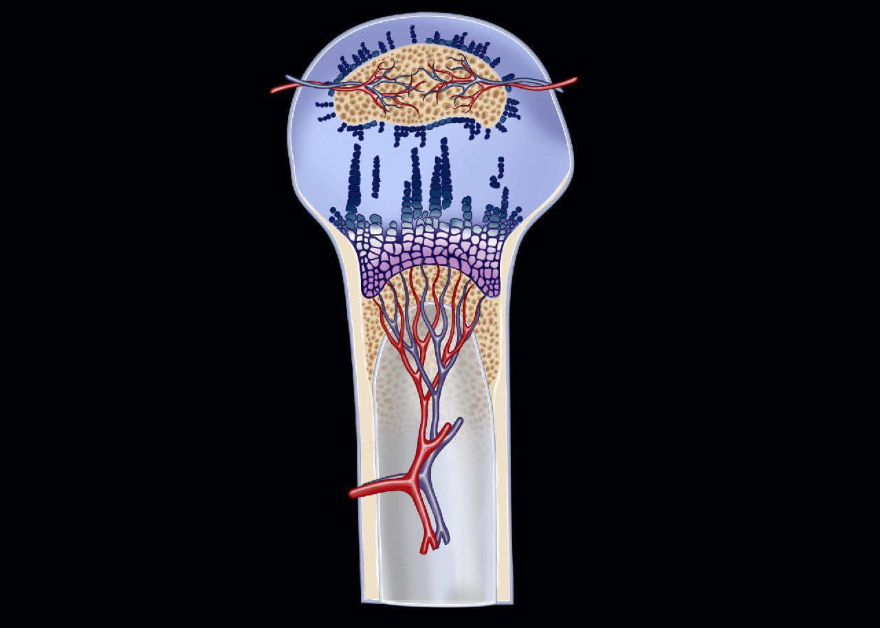
- Hyaline Cartilage Anatomy
- The Physis & Calcification Centers
- Epiphyseal Cartilage
- Fibrocartilage & Hyaline Cartilage
- MR Appearance of Cartilage In Different Age Groups
- FOPE
- Lymphoma of the Bone – 11 min
- Blount Disease
- Gymnast’s Wrist
- Pre-ossification Centers
- Elbow Effusion
- OCD In the Elbow
- Trochlear OCD on MRI
- Trochlear OCD on Arthrogram
- Ultrasound Guided Arthrogram Injection
- OCD In the Capitulum, Loose Body
- Avascular Necrosis in the Elbow
- The Fish Tail Deformity
- OCD In the Knee, LAME
- Legg-Calvé-Perthes disease on X-Ray
- Legg-Calvé-Perthes disease on MRI
- Juvenile Idiopathic Arthritis
- Abscess
- Infection in the Physis
- Tug Lesion
- Salter-Harris Classification System
- Salter-Harris Fracture on X-Ray
- Salter-Harris 2 in the Shoulder
- Salter-Harris 3 in the Knee
- Salter-Harris 3 on CT Imaging
- Indications for MRI in a Pediatric Shoulder
- Performing Arthrograms in the Shoulder
- Ultrasound Guidance in Shoulder Arthrogram
- Salter-Harris 5 on MRI
- Physeal Injury, Cartilage Deformity
- Chondroblastoma in the Knee
- Chondroblastoma in the Ankle
- Anterior Inferior Iliac Spine Avulsion
- Anterior Talofibular Ligament Avulsion
- PCL Tear in a Skeletally Mature Patient
- Jersey Finger
- Cuboid Impaction Fracture
- Femoroacetabular Impingement – 10 min
- Panner Disease
- Osteochondral Lesion of the Radial Head
- Osteochondral Lesion
- Discoid Meniscus
- Discoid Meniscus, Wrisberg Variant
- Plica
- Juvenile Idiopathic Arthritis
- Pigmented Villonodular Synovitis (PVNS)
- Osteomyelitis
- Anorexia Nervosa
- Chondroblastoma
- Chondroblastoma in the Shoulder
- Complex Regional Pain Syndrome
- Lipoblastoma
- Leukemia
- Leukemia, Assessing for Asymmetry
- Myositis Ossificans
- Normal Patchy Bone Marrow
- Osteoblastoma
- Adamantinoma
- Osteoid Osteoma in the Foot
- Osteoid Osteoma in the Finger
- Salter-Harris Classification
- Salter-Harris I Injury
- Salter-Harris II Injury
- Salter-Harris III Injury
- Salter-Harris IV Injury
Content reviewed: September 28, 2021


