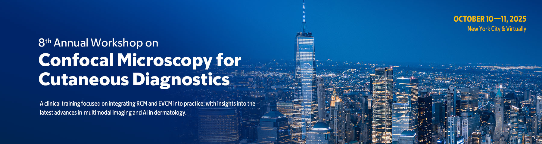
Memorial Sloan Kettering Cancer Center 8th Annual Workshop on Confocal Microscopy for Cutaneous Diagnostics 2025

$50,00
This Product is shared via google drive download link, So please share your correct Gmail id while placing the order .Please note that there are no CME points or certificate associated with this course Samples for Courses Can be found here : Free Samples Here!
Include: 28 videos + 1 pdf, size: 12.7 GB
Related products
PLASTIC SURGERY
$30,00
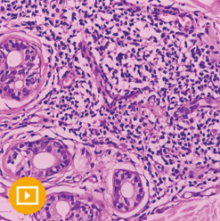
DERMATOLOGY
$35,00
DERMATOLOGY
$35,00

DERMATOLOGY
$35,00

$20,00
DERMATOLOGY
$10,00