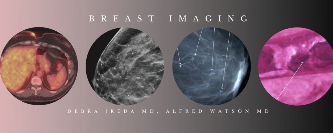
CME Science Breast Imaging BUNDLE (Programs 1-3) Debra Ikeda MD, Alfred Watson MD 2020
$100,00
Samples for Courses Can be found here : Free Samples Here!
CME Science Breast Imaging BUNDLE (Programs 1-3) Debra Ikeda MD, Alfred Watson MD 2020
CME Science Breast Imaging BUNDLE (Programs 1-3) Debra Ikeda MD, Alfred Watson MD 2020
CME Science Breast Imaging BUNDLE (Programs 1-3) Debra Ikeda MD, Alfred Watson MD 2020 Dr. Watson is a Distinguished Emeritus Professor of Radiology at Baylor College of Medicine and a Fellow in the Society of Breast Imaging. Dr. Watson has held the positions of Colonel USAF 1969-1984; Aeronautical Rating Chief Flight Surgeon; Chief Aerospace Medicine, Headquarters USAF Europe Office of Surgeon General, 1973-1975; Military Consultant to the USAF Surgeon General for Radiology
home page : https://cmescience.teachable.com/p/breast-imaging-bundle-debra-ikeda-m-d-alfred-watson-m-d
CME Science Breast Imaging (BUNDLE) – Debra Ikeda M.D., Alfred Watson M.D 2020
YOU WILL GET THE COURSE VIA Google Drive DOWNLOAD LINK (FAST SPEED) AFTER PAYMENT
Debra Ikeda, M.D.
• Professor of Radiology, Breast Imaging
• Stanford University School of Medicine
Dr. Ikeda is a tenured Professor of Radiology at Stanford and was Breast Imaging Section Chief from 1992-2016. She has published over 110 scientific articles, led (2004) or was vice-chair (2013) of the ACR BI-RADS-MRI Committee, and authored “Breast Imaging: The Requisites 3rd Edition” (2016). She is a recognized breast imaging/biopsy speaker/teacher, with over 300 presentations in the USA and around the world. Prior research focused on FFDM/DBT, needle biopsy, MRI/MRI biopsy, compliance/outcomes, density legislation. The new research involves mammographic positioning, DWI, breast cancer/stroma genetics, MRI outcomes, DBT interval cancers.
Alfred B. Watson Jr. MD MPH FACR FACPM
- Distinguished Emeritus Professor of Radiology
- Baylor College of Medicine
- Fellow of the American College of Radiology
Dr. Watson is a Distinguished Emeritus Professor of Radiology at Baylor College of Medicine and a Fellow in the Society of Breast Imaging. Dr. Watson has held the positions of Colonel USAF 1969-1984; Aeronautical Rating Chief Flight Surgeon; Chief Aerospace Medicine, Headquarters USAF Europe Office of Surgeon General, 1973-1975; Military Consultant to the USAF Surgeon General for Radiology, 1979-1984. As a member of Breast Imaging Training Advisory Committee, he helped to develop and publish the breast imaging residency and fellowship training curriculum.
Dr Watson has delivered over 148 invited lectures nationally and 9 international visiting professor assignments at academic and military programs. He has delivered over 1,000 hours of CME breast lectures for residents, breast fellows, radiologists and Clinicians. Dr Watson was selected by the ABR as an Oral Board examiner in Breast Imaging 16 of the last 19 years. For his commitment and excellence, he was awarded Distinguished Service Award by the ABR in 2008, 2010 and 2014. The crowning landmark came in 2015 when ABR honored him with a Lifetime Achievement Award. In his 37-year career with the US Air Force and Baylor College of Medicine, Dr. Watson trained generations of physicians with unwavering energy and enthusiasm.
Release date: September 16, 2020
Estimated time to complete activity: 17 hours
Educational Objectives
At the conclusion of this activity, participants should better be able to:
- Optimize imaging methods to reduce the number of false-positive mammography interpretations.
- Describe recent advances and techniques in breast imaging.
- Integrate information presented in this course into efforts to improve the imaging skills of the participants.
Target Audience
This course is intended for Practicing Radiologists and Radiologic Nurses, Physician Assistants, Technologists, Scientists, Residents, Fellows and others who are interested in current techniques and applications for breast imaging.
Topics/Speakers :
Debra Ikeda, M.D. – Program 1
- Update on MRI Screening and Diagnosis
(37:21) - Update on Digital Breast Tomosynthesis Screening and Diagnosis
(43:03) - Wireless Localization Techniques on Mammo, US and Stereo
(53:12) - Multimodality (Mammo/US/MRI) Breast Imaging Cases
(56:52) - Ultrasound and Stereotactic Core Biopsy
(42:50) - Augmented/Reconstructed Breast with Lipofilling and the New Faces of Fat Necrosis on Mammo, US and MRI
(55:20) - Mammography Legislation, Breast Density Legislation and Risk
(38:29)
Alfred Watson, M.D. – Program 2
-
Imaging Breast Calcifications and Review of Benign Calcifications (Part 1)
(34:54) - Imaging Breast Calcifications and Review of Benign Calcifications (Part 2)
(32:39) - Imaging Evaluation and Discussion of Breast Asymmetry
(64:04) - Morphology Criteria for Malignant Breast Calcifications (Part 1)
(30:45) - Morphology Criteria for Malignant Breast Calcifications (Part 2)
(30:24) - The Male Breast – Imaging, Diagnosis, and Treatment of Male Breast Disease
(76:48) - Work-Up of Palpable and Non-Palpable Mass(es)Work-Up of Palpable and Non-Palpable Mass(es) (Part 1)
(41:55) - Work-Up of Palpable and Non-Palpable Mass(es)Work-Up of Palpable and Non-Palpable Mass(es) (Part 2)
(45:11)
Alfred Watson, M.D. – Program 3
- Review Breast Anatomy and Ductal Pathology with Imaging Correlation
(58:18) - Interesting Breast Imaging Cases with Teaching Points
(74:45) - Review Breast Lobular Pathology with Mammogram and Ultrasound Imaging Correlation
(53:25) - Risk Management for Breast Imagers
(101:40)
Related products
OBSTETRICS & GYNECOLOGY
OBSTETRICS & GYNECOLOGY
GCUS Ultrasound Applications in Austere/Rural Medicine 2020 (VIDEOS)
ENDOCRINE / NUTRITION
UCSF Obstetrics and Gynecology Update: What Does The Evidence Tell Us 2023
OBSTETRICS & GYNECOLOGY
AIUM Fetal Abdominal and Thoracic Abnormalities: Features and Diagnosis

