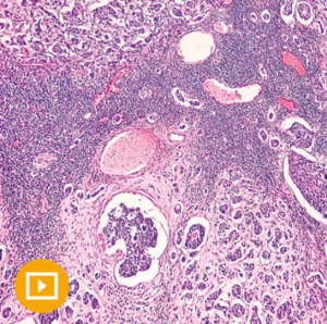
Breast Pathology 2023
$35,00
24 videos + 2 pdfs, size: 7.14 GB
Contact me
This Product is shared via google drive download link, So please share your correct Gmail id while placing the order .Please note that there are no CME points or certificate associated with this course
Breast Pathology 2023 — from Oakstone CME’s Masters of Pathology Series — outlines best practices for specimen handling and reporting, defined diagnostic criteria, improved recognition of less common diagnostic entities, and accurate interpretation of ancillary studies by using both routine microscopic examination and immunohistochemistry.
Directed by Laura C. Collins, MD, this breast pathology continuing medical education will help define the role of newer adjunctive molecular tests and will also help you to better:
- Classify proliferative breast, in situ, papillary, fibroepithelial, spindle cell, and vascular lesions
- Make the distinction between invasive and in situ lesions
- Identify a variety of uncommon benign and malignant lesions
- Understand guidelines for reporting prognostic and predictive factors
Date of Original Release: March 1, 2023
Date Credits Expire: February 28, 2026
CME credit is awarded upon successful completion of a course evaluation and post-test.
This activity meets the American Board of Pathology’s (ABPath) Continuing Certification program requirements for Part II (CME) Lifelong Learning.
Learning Objectives
At the conclusion of this CME activity, the participant will be able to:
- Evaluate diagnostic criteria and clinical significance of various common and uncommon benign, in situ, and malignant lesions of the breast in both core needle biopsy specimens and surgical excision specimens
- Describe differential diagnostic problems commonly encountered in breast pathology and develop strategies to resolve them in the practice environment
- Utilize the latest information on the uses and limitations of immunohistochemistry in resolving diagnostic problems in breast pathology
- Understand the updated guidelines for ER, PR, and HER2 testing and reporting
- Understand the applications of molecular testing in breast cancer
Intended Audience
This educational activity was designed for practicing pathologists, pathology assistants, and residents.
24 videos + 2 pdfs, size: 7.14 GB
Topics:
- 1. Intraductal Proliferative Lesions.mp4
2. Intraductal Proliferative Lesions – Microscopy Session.mp4
3. Papillary Lesions of the Breast.mp4
4. Papillary Lesions of the Breast – Microscopy Session.mp4
5. Contemporary Considerations for Breast Core Needle Biopsies.mp4
6. Lobular Carcinomas of the Breast.mp4
7. Breast Fibroepithelial Tumours – An Update.mp4
8. Breast Fibroepithelial Tumours – Slide Seminar.mp4
9. Mucinous Lesions of the Breast.mp4
10. Mucinous Lesions of the Breast – Microscopy Session.mp4
11. Spindle Cell Lesions of the Breast.mp4
12. Spindle Cell Lesions of the Breast – Microscopy Session.mp4
13. Vascular Lesions of the Breast.mp4
14. Vascular Lesions of the Breast – Microscopy Session.mp4
15. Epithelial-Myoepithelial Lesions of the Breast.mp4
16. Epithelial-Myoepithelial Lesions of the Breast – Microscopy Session.mp4
17. Small Glandular Proliferations & Mimics of Breast Carcinoma.mp4
18. Small Glandular Proliferations – Microscopy Session.mp4
19. Inflammatory & Reactive Conditions in the Breast.mp4
20. The Spectrum of Triple Negative Breast Cancers.mp4
21. The Spectrum of Triple Negative Breast Cancers – Microscopy Session.mp4
22. Breast Pathology in the Era of Genomics.mp4
23. Updates in Immunohistochemistry in Diagnostic Breast Pathology.mp4
24. Applications of Digital Pathology in the Field of Breast Pathology.mp4
Questions.pdf
Syllabus.pdf
Related products
CRITICAL CARE / EMERGENCY MEDICINE
OBSTETRICS & GYNECOLOGY
OBSTETRICS & GYNECOLOGY
AIUM Fetal Abdominal and Thoracic Abnormalities: Features and Diagnosis
CRITICAL CARE / EMERGENCY MEDICINE
The Brigham Board Review in Infectious Diseases Brigham and Women’s Hospital Board Review
HARVARD MEDICINE

