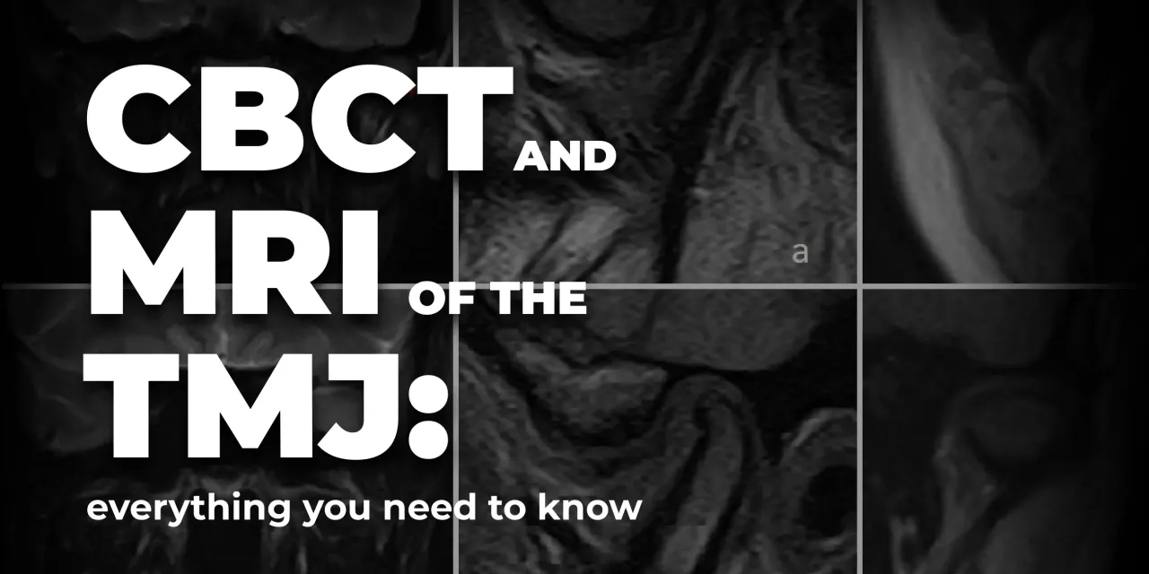
OHI-S CBCT and MRI of the TMJ everything you need to know

$50,00
This Product is shared via google drive download link, So please share your correct Gmail id while placing the order .Please note that there are no CME points or certificate associated with this course Samples for Courses Can be found here : Free Samples Here!
+ Include: 4 videos + 4 audios + 4 file sub vtt, size: 2.39 GB
+ Include: 4 videos + 4 audios + 4 file sub vtt, size: 2.39 GB
+ Target Audience: orthodontists, prosthodontists, functional dentists, general dentists
+ Information:
4 lessons (4h 9min)
Acquire TMJ imaging protocols using modern methods such as CBCT and MRI from leading experts in the field: Mariano Rocabado, Lukas Lassmann and Kaan Orhan!
You will first learn the anatomy of the TMJ, as this topic is fundamental. Then, the lecturers will teach you:
– Diagnostic algorithms and stages of MR examination of the TMJ
– Dr. Mariano Rocabado’s CBCT protocols
– Diagnosis of pathologies such as сortical bone erosion, idiopathic condyle resorption, subcortical cysts, TMJ ankylosis, developmental defects of the TMJ, displacement of the disc.
This course will serve as your guide to modern TMJ imaging methods!
The OHI‑S CBCT and MRI of the TMJ: Everything You Need to Know course is best for prosthodontists, oral surgeons, orthodontists, and radiologists who want structured, training in imaging protocols for temporomandibular joint (TMJ) diagnosis and treatment planning. It emphasizes evidence‑based use of CBCT and MRI to evaluate joint anatomy, pathology, and function.
 Who Should Enroll
- Prosthodontists managing occlusal rehabilitation and TMJ disorders in restorative cases.
- Oral & maxillofacial surgeons diagnosing and treating surgical TMJ conditions.
- Orthodontists integrating TMJ imaging into treatment planning for malocclusions and functional corrections.
- Radiologists & dental imaging specialists interpreting CBCT and MRI scans of the TMJ.
- Residents & fellows in prosthodontics, orthodontics, oral surgery, or radiology seeking structured TMJ imaging training.
 What You’ll Learn
- CBCT protocols: indications, scanning techniques, and interpretation of bony structures.
- MRI protocols: soft tissue evaluation, disc position, and joint function analysis.
- Comparative imaging: when to use CBCT vs. MRI for specific TMJ conditions.
- Diagnostic accuracy: identifying pathology such as disc displacement, degenerative changes, and inflammatory conditions.
- Clinical integration: applying imaging findings to treatment planning and patient management.
- Case‑based lessons: real examples demonstrating CBCT and MRI use in TMJ diagnosis.
+ Topics:
Lesson 1.Clinical anatomy of the Synovial Temporomandibular Joint
– Physiological anatomy of TMJ movement
– CBCT analysis of the TMJ. Open and closed mouth
– Evaluation of normal TMJ and clinical cases of TMD
– Osteochondritis dissecans and avascular necrosis
– CBCT and MRI considerations in various clinical cases.
Recommended for: Orthodontists, Prosthodontists, Functional dentists, General dentists.
Lesson 2.MRI of TMJ
– Timeline of MRI imagining
– Fundamentals of MRI examination
– Mechanism of MRI machine function. The procedure
– TMJ MRI basic anatomy
– TMJ disc and structure anatomy
– Patterns of basic pathologies
– MRI of muscles
– Connections with TMJ and temporal bone and surrounding tissues.
Recommended for: Prosthodontists, Implantologists, Oral and maxillofacial surgeons, Functional dentists, Therapeutists, Endodontists, Pediatric dentists, Orthodontists, General dentists.
Lesson 3.Cone Beam Computed Tomography Protocols of Dr. Mariano Rocabado. Part 1
– Analysis of cranioverteral according to Dr. Mariano Rocabado
– Normal occiput atlas axis flexion
– Craniofacial assymetry evalaution
– CBCT and x-ray static and dynamic cervical and TMJ protocol
– Functional analysis by Dr. Mariano Rocabado
– Step-by-step of clinical analysis of various cases.
Recommended for: Orthodontists, Prosthodontists, Functional dentists, General dentists.
Lesson 4.Cone Beam Computed Tomography Protocols of Dr. Mariano Rocabado. Part 2
– Anatomy: what you need to know for the treatment of TMJ pathology
– Tricentric approach: advantages and special features
– Map of joint pain: areas of sensitivity in the joint
– Assessment of the state of the TMJ by CBCT
– Static assessment of the spine from the side
– Functional analysis by Dr. Mariano Rocabado
– Craniocervical mandibular (CCM) tricentric relation
– Step-by-step clinical analysis of various cases.
Recommended for: Orthodontists, Prosthodontists, Functional dentists, General dentists.
Related products